Home > Animals > Mammals > Muridae > Water Mouse
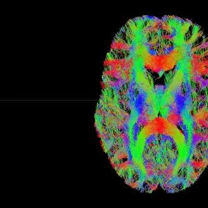
White matter fibres, brain mri scan C014 / 5674

![]()
Wall Art and Photo Gifts from Science Photo Library
White matter fibres, brain mri scan C014 / 5674
White matter fibres overlaid a mri scan of the human head. Coloured 3D diffusion spectral imaging (DSI) scan of the bundles of white matter nerve fibres in the brain. The fibres transmit nerve signals between brain regions and between the brain and the spinal cord. Diffusion spectrum imaging (DSI) is a variant of magnetic resonance imaging (MRI) in which a magnetic field maps the water contained in neuron fibers, thus mapping their criss-crossing patterns. A similar technique called diffusion tensor imaging (DTI) is also used to explore neural data of white matter fibres in the brain. Both methods allow mapping of their orientations and the connections between brain regions. Data/software: NIH Human Connectome Project /humanconnectomeproject.org)
Science Photo Library features Science and Medical images including photos and illustrations
Media ID 9224189
© PASIEKA/SCIENCE PHOTO LIBRARY
Brain Scan Connections Diffusion Spectral Imaging Diffusion Tensor Imaging Dsi Scan Dti Scan Fibers Fibre Tracking Fibres Human Brain Magnetic Resonance Imaging Mapping Mri Scan Nerve Bundles Nerve Fibre Pathway Pathways White Matter Nervous System
FEATURES IN THESE COLLECTIONS
> Animals
> Mammals
> Muridae
> Water Mouse
> Maps and Charts
> Related Images
EDITORS COMMENTS
This print showcases the intricate network of white matter fibres in the human brain, overlaid on an MRI scan. The coloured 3D diffusion spectral imaging (DSI) scan beautifully reveals the bundles of nerve fibres that transmit crucial signals between different regions of the brain and even to the spinal cord. Utilizing a magnetic field to map water within neuron fibers, DSI allows for a detailed visualization of their criss-crossing patterns. This technique is part of the broader field of magnetic resonance imaging (MRI), specifically tailored to explore white matter pathways in our nervous system. Another similar method called diffusion tensor imaging (DTI) is also employed to study neural data related to these white matter fibres. Both DSI and DTI enable researchers and medical professionals to map not only the orientations but also uncover connections between various brain regions. These invaluable insights into our complex neural architecture contribute significantly towards understanding how information flows throughout our brains. The data used for this remarkable image originates from NIH Human Connectome Project, an ambitious endeavor aiming to comprehensively map human brain connectivity. By shedding light on these intricate pathways and connections, studies like this pave the way for advancements in neuroscience research, diagnostics, and potentially even therapeutic interventions. Captured by PASIEKA/SCIENCE PHOTO LIBRARY, this visually stunning print serves as a testament to both scientific progress and artistic beauty found within our own minds.
MADE IN THE USA
Safe Shipping with 30 Day Money Back Guarantee
FREE PERSONALISATION*
We are proud to offer a range of customisation features including Personalised Captions, Color Filters and Picture Zoom Tools
SECURE PAYMENTS
We happily accept a wide range of payment options so you can pay for the things you need in the way that is most convenient for you
* Options may vary by product and licensing agreement. Zoomed Pictures can be adjusted in the Cart.